Understanding the Pelvis Sacrum Relationship: Anatomy, Function, and Lifelong Care
- Updated - January 11, 2025
The human body’s structural integrity depends upon a finely tuned interplay of bones, joints, muscles, and connective tissues. At the heart of this dynamic system is the pelvis sacrum relationship.
Though often overshadowed by more “glamorous” joints like the hips or shoulders, the subtle synergy between the bony pelvis and the sacrum quietly anchors posture stabilizes movement and provides a bridge between the spine and the lower limbs.
By understanding how these structures work together—and how their relationship evolves over a lifetime—we can gain insights into preventing pain, enhancing performance, and living with greater ease.
Introduction to the Pelvis and Sacrum
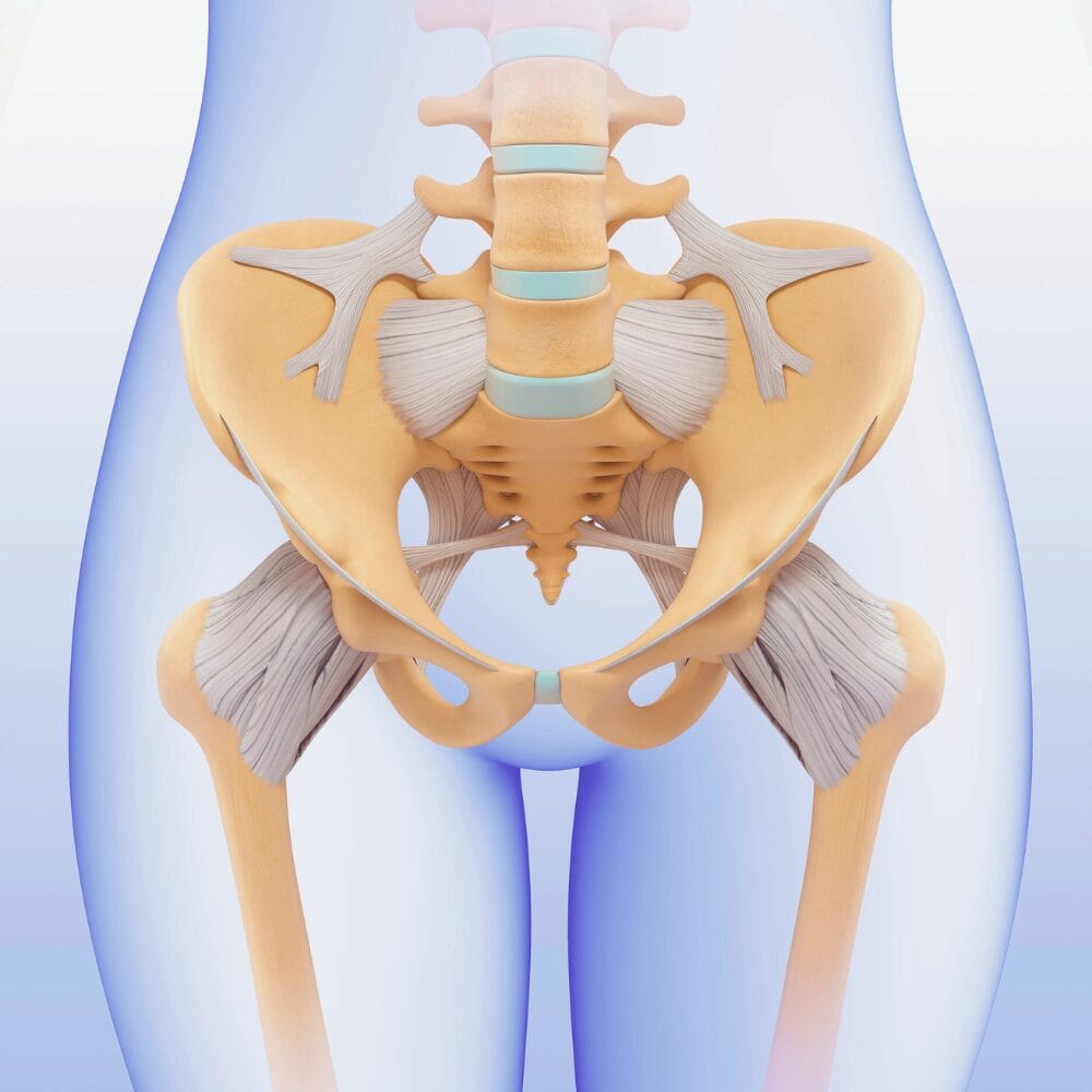
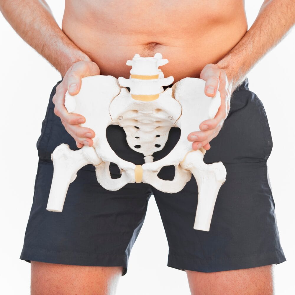
The pelvis is a complex bony structure that forms the base of the spine and the floor of the abdominal cavity. It comprises four bones: the ilium, ischium, pubis, and sacrum. The sacrum, a large, triangular bone at the base of the spine, is formed by the fusion of five sacral vertebrae (S1–S5) between the ages of 18 and 30. This fusion process creates a solid, supportive structure that is crucial to the body’s stability and movement.
The pelvis and sacrum provide a sturdy foundation for the upper body, facilitating movement and protecting the pelvic organs. The sacrum articulates with the pelvic bones at the sacroiliac joints, which, although allowing only minimal movement, are essential for load distribution and shock absorption. This intricate relationship between the pelvis and sacrum ensures that the body can perform a wide range of activities, from walking and running to lifting and bending, with stability and efficiency.
Anatomy at a Glance
The pelvis comprises two large hip bones (the ilia) fused with the ischium and pubis to form a ring. At the back of this pelvic ring sits the sacrum, a wedge-shaped bone formed by the fusion of five sacral vertebrae. The sacrum articulates with the pelvic bones at the sacroiliac (SI) joints, where only minimal movement occurs. Below the sacrum, the coccyx completes the structure at the base of the spine.
The superior articular processes of the first sacral vertebra articulate with the inferior articular processes of the fifth lumbar vertebra, emphasizing the importance of this junction for spinal structure and function.
This pelvis–sacrum unit supports the upper body’s weight and transfers forces into the legs. Although each component has unique characteristics, they work as a cohesive system essential for walking, running, lifting, and maintaining a stable, upright posture.
Discover a practitioner near you.
Looking for a practitioner near you? Our extensive network of qualified professionals is here to help you.
The Sacroiliac Joint: A Subtle but Vital Connection

The SI joints, where the sacrum meets the ilium on each side, are not designed for large ranges of motion. Instead, they permit only small gliding movements—seemingly insignificant, yet critical for load distribution, shock absorption, and core stability.
The acetabulum of the pelvis articulates with the proximal femur at the hip joint, highlighting its functional importance in transferring forces and stabilizing the musculoskeletal system.
Key Functions of the SI Joint:
- Force Transmission: The SIJs channel forces from the upper body through the sacrum into the pelvis and down the legs. Slight misalignments or stiffness here can alter how loads are distributed, contributing to discomfort or instability.
- Shock Absorption: Subtle SIJ motions help absorb impact, which is important during activities like running or jumping. Other joints may compensate without this, potentially leading to injury over time.
- Spinal Stability: Though the SIJs move only slightly, they serve as crucial stabilizers. Robust ligaments and balanced muscle activation keep these joints strong and secure, reducing stress on the spine.
Pelvic Girdle Positioning and Posture
The orientation of the sacrum relative to the pelvis influences the entire spine. Nutation (forward tilt of the sacrum) and counternutation (backward tilt) are small but meaningful movements that affect lumbar lordosis and overall posture.
- Nutation: Promotes a slight increase in the lumbar curve and occurs naturally in movements like bending forward or squatting.
- Counternutation: Reduces the lumbar curve and may appear when leaning backward or compensating for particular pelvic tilts.
Pelvic floor dysfunction can lead to contraction of the pelvic floor muscles when relaxation is required, complicating posture and movement.
A healthy balance between nutation and counternutation supports proper spinal alignment, muscle activation patterns, and efficient force distribution.
The Role of Musculature and Posterior Sacroiliac Ligaments
A harmonious pelvis–sacrum relationship relies on a network of muscles, fascia, and ligaments:
- Pelvic Floor Muscles: Forming a supportive “hammock,” they influence pelvic stability, bladder and bowel control, and core function.
- Deep Core Muscles (Transversus Abdominis, Multifidus): These muscles stabilize the spine and pelvis from within, aiding in SI joint stability and evenly distributing forces.
- Hip and Gluteal Muscles: Strong glutes and well-coordinated hip muscles ensure proper pelvic positioning. Weakness or imbalance here can shift stress onto the sacrum and spine.
- Thoracolumbar Fascia and Ligaments: Connective tissues maintain tension and alignment. They help keep the pelvis and sacrum properly oriented when balanced and supple.
The posterior sacroiliac ligament provides attachments within the sacrum and contributes to the stability of the sacral vertebrae.
Pelvis and Abdominal Cavity Relationship
The pelvis and abdominal cavity are intricately connected, with the pelvis forming the floor of the abdominal cavity. The pelvic cavity, a space enclosed by the pelvic bones, houses critical organs such as the urinary bladder, pelvic colon, internal reproductive organs, and rectum. This cavity is superior to the perineum, which lies below the pelvic floor. The pelvic floor is a muscular layer that separates the pelvic cavity superiorly from the perineum, providing essential support and maintaining the integrity of the pelvic organs.
The pelvic cavity functions as a protective chamber, safeguarding the organs within it from external forces and maintaining their proper positioning. This relationship between the pelvis and abdominal cavity is vital for the overall health and functionality of the body’s lower regions. By understanding this connection, we can better appreciate the importance of maintaining pelvic health and stability throughout our lives.
Embryology and Development
The development of the pelvis is a fascinating process that begins in the embryonic stage. The ilium, ischium, and pubis bones originate from the lateral plate mesoderm, while the sacrum forms from the fusion of five sacral vertebrae. This development occurs in a cephalocaudal direction, meaning it progresses from the head downwards. The ilium and ischium are the first to form, followed by the pubis bone, with the sacrum developing last.
Genetic and environmental factors play significant roles in the development of the pelvis. Any abnormalities during this process can lead to congenital malformations, which may affect the structure and function of the pelvis and sacrum. Understanding the embryological development of these structures helps us appreciate the complexity and precision required for their proper formation. It highlights the importance of early detection and intervention in cases of developmental abnormalities.
Physiology
The pelvis is a cornerstone of the body’s movement and support systems. It plays a crucial role in activities such as walking, running, and climbing stairs by working in concert with the sacroiliac joints to decrease the force transferred from the ground and lower extremities to the spine and upper body. This force distribution is essential for maintaining balance and preventing injury.
The pelvic floor muscles, including the levator ani and coccygeus muscles, provide additional support to the pelvic organs and help maintain continence. These muscles form a supportive “hammock” that influences pelvic stability and core function. The pelvic cavity additionally houses vital organs such as the urinary bladder, pelvic colon, internal reproductive organs, and rectum, underscoring its importance in protecting and supporting these structures.
Pelvic connective tissue, including the posterior sacroiliac ligaments and lateral sacral arteries, further enhances the stability and functionality of the pelvis. These tissues provide structural support and ensure proper alignment, contributing to the overall health and efficiency of the pelvis and sacrum. By understanding the physiological functions of the pelvis, we can better appreciate its role in maintaining our body’s stability and movement throughout our lives.
Lifespan Perspectives: Changes Over Time
Childhood and Adolescence: During growth and development, bones are still fusing, and alignment patterns are being established. Balanced activities and encouraging symmetrical movement patterns can set the stage for a stable pelvis–sacrum relationship later in life. The pubic bone provides critical muscle attachment points and contributes to overall stability within the pelvic girdle.
Adulthood: In the prime of life, everyday habits, athletic pursuits, and work demands shape pelvic alignment. Maintaining good posture, incorporating regular exercise, and managing muscle imbalances are key to preserving SIJ health and preventing pain.
Older Adulthood: As we age, bone density may decrease, and degenerative changes can affect joints. Gentle stability and mobility exercises, attention to posture, and interventions like manual therapy can help preserve comfort, reduce stiffness, and maintain function over time.
Gender Differences and Considerations
The female pelvis tends to be wider and shaped differently to support childbirth. Hormonal fluctuations during pregnancy, postpartum, and menopause can affect ligamentous laxity and pelvic floor integrity, influencing the SI joints. Pregnancy-related hormones like relaxin increase joint mobility, while changes in the center of gravity and pelvic floor stress require postpartum re-stabilization. The internal iliac artery plays a crucial role in supplying oxygenated blood to various pelvic organs such as the bladder, prostate, uterus, and vagina. Tailored exercises and professional guidance can greatly assist women in maintaining pelvic health through life’s transitions.
Integrating with Other Bodily Systems
The pelvis–sacrum relationship does not exist as an isolated structure; it functions within a complex, whole-body matrix of bones, muscles, connective tissues, and neural feedback loops. Understanding how the pelvis and sacrum interact with other systems helps illuminate why localized dysfunction can have far-reaching consequences and how addressing these influences can lead to more lasting and comprehensive improvements in health and performance.
The median sacral artery is crucial in supplying blood to the sacral region, highlighting its importance in sacral anatomy alongside structures such as the sacral nerves and rectum.
Respiratory Diaphragm Interaction:
The diaphragm, situated beneath the rib cage, and the pelvic floor at the base of the pelvis form the top and bottom of the body’s core canister. Together, they help manage intra-abdominal pressure, influencing spinal stability and posture.
When the diaphragm contracts and relaxes rhythmically with each breath, it coordinates with the pelvic floor, transversus abdominis, and multifidus muscles to maintain a stable yet dynamic core. The posterior sacral foramina serves as passageways for the sacral nerve fibers and is structurally significant, being smaller than the anterior sacral foramina, which provides entry and exit points for nerve divisions associated with the sacral canal. If the diaphragm’s function is compromised—perhaps due to shallow chest breathing, poor posture, or stress—the pelvic floor often responds by altering its tension.
Over time, this can shift the sacrum’s orientation, affect how loads pass through the SI joints, and influence low back comfort. Incorporating diaphragmatic breathing exercises, breath-focused yoga, or mindful relaxation techniques can help restore a healthy interplay between these structures, indirectly supporting sacral positioning and improving SI joint stability.
Lower-Limb Biomechanics:
The pelvis–sacrum relationship is also molded by what happens below the waist. Each step we take involves a complex transfer of forces through the feet, ankles, knees, hips, and into the pelvis.
When alignment issues occur in the lower limbs—such as overpronation (the foot rolling inward excessively), supination (foot rolling outward), or knee valgus (knees collapsing inward)—the repercussions can travel upward. For instance, overpronation alters the angle at which force enters the leg, potentially rotating the tibia and femur in ways that shift pelvic alignment. This subtle chain reaction can tilt or rotate the sacrum, increasing stress on the SI joints and lower spine.
The pelvic girdle connects various bones and joints, supports movement, and provides stability. By addressing foot mechanics through proper footwear, orthotics, or targeted foot and ankle exercises, and by strengthening and stabilizing the hip girdle, we can help ensure a balanced foundation that supports a healthy pelvis–sacrum relationship.
Upper Body and Shoulder Girdle:
Just as issues in the lower limbs can migrate upward, misalignments and muscular imbalances in the upper body can cascade downward. The thoracic spine, rib cage, and shoulder girdle all contribute to the body’s overall postural balance. If the shoulders are rounded forward, the head juts ahead of the spine, or the thoracic spine becomes excessively kyphotic (hunched), the body often compensates by adjusting the position of the pelvis. Over time, these compensations can alter sacral orientation, change muscle firing patterns in the trunk and hips, and affect SI joint stability. For example, a stiff upper back may cause the lumbar spine and pelvis to tilt in ways that impose uneven stress on the sacroiliac joints. Restoring proper thoracic mobility, strengthening the scapular stabilizers, and refining head and neck alignment can minimize these downward compensations. Activities like corrective exercise programs, Pilates, swimming, or working with a postural specialist can help achieve a more harmonious upper-lower body interplay, thereby fostering a more stable and balanced pelvis–sacrum relationship.
By recognizing the interconnectedness of the respiratory, lower-limb, and upper-body systems, we better understand why pelvis–sacrum health cannot be addressed in isolation. Each system influences the others, and imbalances in one area can create repercussions throughout the entire kinetic chain. Embracing this holistic perspective enables more effective prevention and intervention strategies — ensuring that any efforts to improve pelvic and sacral function also support the whole person’s health and well-being.
Common Issues and Beyond Exercise: Clinical Interventions for Sacroiliac Joint Dysfunction
Sacroiliac Joint Dysfunction: When SIJ movement becomes restricted, inflamed, or unstable, lower back or buttock pain often results. As a complex bony structure, the bony pelvis plays a crucial role in load-bearing and stability, and issues here can significantly impact overall pelvic function. Targeted exercises, manual therapy, chiropractic care, or osteopathic treatments can help restore balance and alleviate discomfort. SI belts offer temporary support, while techniques like myofascial release, neuromuscular re-education, and acupuncture may contribute to lasting relief.
Exercise Considerations
Start with Stabilization: Begin by strengthening the core and pelvic floor. Planks, bird-dog exercises, and gentle pelvic tilts help establish a neutral sacrum and stable foundation.
Focus on Gluteal and Hip Strengthening: Glute bridges, clamshells, lateral band walks, and hip thrusts build strong, aligned hips that support SIJ stability. Strengthening the muscles around the hip joint is crucial for maintaining SIJ stability, as the hip joint plays a key role in transferring forces and stabilizing the musculoskeletal system.
Mindful Mobility: Gentle hip openers, hip flexor stretches, and balanced hamstring lengthening exercises prevent excessive pelvic tilts and maintain healthy sacral positioning.
Functional Movement Patterns: Exercises mirroring daily activities—squats, lunges, hip hinges—reinforce proper alignment and help translate stability into everyday life.
Listen to Your Body: If specific movements cause pain, scale back, modify the range of motion or seek professional advice. Addressing issues early prevents chronic dysfunction.
Preventative Strategies and Lifestyle Factors
Optimize Posture and Ergonomics: Sitting upright, adjusting workstations, and avoiding prolonged static postures maintain a neutral pelvis and reduce SIJ stress.
Footwear and Grounding: Supportive shoes and mindful foot placement influence force distribution through the legs and pelvis.
Balanced Activities and Rest: A mix of movement and rest, adequate sleep, and stress management support tissue health and better overall alignment.
Nutrition and Bone Health: Calcium, vitamin D, protein intake, and weight-bearing exercise support bone density and ligamentous integrity, preserving pelvic stability over time.
Maintaining the health of the internal iliac artery is crucial for overall pelvic health, as it supplies oxygenated blood to various pelvic organs such as the bladder, prostate, uterus, and vagina.
Final Thoughts
The pelvis and sacrum form a quiet yet powerful partnership that serves as the body’s central hub of stability, posture, and movement. By appreciating how these structures interact, adapting to changes throughout the lifespan, considering gender-specific factors, and integrating clinical and complementary approaches alongside thoughtful exercise and lifestyle strategies, we create a more holistic understanding of pelvic health.
A balanced pelvis–sacrum relationship doesn’t just relieve pain — it can enhance athletic performance, improve posture, and allow us to move through life with greater comfort and confidence. Embracing this integrated perspective provides a foundation for long-term musculoskeletal well-being and resilience, regardless of age or stage of life.
Resources
Articles:
The Sacroiliac Joint: An Overview of Its Anatomy, Function, and Potential Clinical Implications
Journal of Anatomy, 2012.
This comprehensive review examines the anatomy and function of the sacroiliac joint, discussing its role in load transfer between the spine and lower limbs and its relevance in conditions like low back pain.
Age-Related Changes in the Articular Cartilage of Human Sacroiliac Joint
Anatomy and Embryology, 1998.
This study investigates how aging affects the articular cartilage of the sacroiliac joint, providing insights into degenerative changes that can impact joint function over time.
Gender Differences in Unintended Anterior Pelvic Roll During Primary Hip Arthroplasty
Journal of Orthopaedic Surgery and Research, 2024.
This research explores how pelvic morphology differs between genders and the clinical significance of these differences, particularly in surgical contexts.
Sacroiliac Joint Dysfunction: Pathophysiology, Diagnosis, and Treatment
European Spine Journal, 2021.
This article reviews the anatomy and function of the sacroiliac joint, as well as the pathophysiology, clinical presentation, diagnostic criteria, and treatment options for sacroiliac joint dysfunction.
PLEASE NOTE
PostureGeek.com does not provide medical advice. This information is for educational purposes only and is not intended to be a substitute for professional medical attention. The information provided should not replace the advice and expertise of an accredited health care provider. Any inquiry into your care and any potential impact on your health and wellbeing should be directed to your health care provider. All information is for educational purposes only and is not intended to be a substitute for professional medical care or treatment.
About the author
Join our conversation online and stay updated with our latest articles.
Find Expert Posture Practitioner Near You
Discover our Posture Focused Practitioner Directory, tailored to connect you with local experts committed to Improving Balance, Reducing Pain, and Enhancing Mobility.